Anatomy of ovaries
The ovaries are the two female reproductive glands which are solid, pinkish grey and almold-shape entities situated on either side of the uterus and are connected to the uterus by the fallopian tube (fig. 1). The right ovary tends to be slightly larger than the left ones

Fig. 1 (Copyright © 2008 by MediScene) The female reproductive organs
The ovaries are suspended within the peritoneal cavity by a fold of peritoneum called the mesovarium (a subdivision of broad ligament of uterus). The proximal edge of the ovary is connected to the lateral angle of the uterus by another ligament called the ligament of ovary (fig. 2)
The suspensaory ligament provides a less supportive function in the sense that, it carries vessels, nerves and lymphatic to and fro the ovaries.
The peritoneum only hold (support) the ovaries, but it does not cover it as some may expect, and therefore the eggs released during the ovulation passes into the peritoneal cavity.

Fig. 2 (Copyright © 2010 by Wolter Kluwer); Ligaments supporting the ovaries, uterus and the uterine tube (Moore et al., 2010)
The ovaries are located within the true pelvis (fig. 3), but during pregnancy, they are lifted out of the true pelvis due to uterine enlargement. Similarly, the premenopausal ovaries are larger than the postmenopausal ovaries. The postmenopausal ovaries shrink and atrophy when the graafian follicles and ova disappear and become an inert residue consisting of white connective tissue.
.
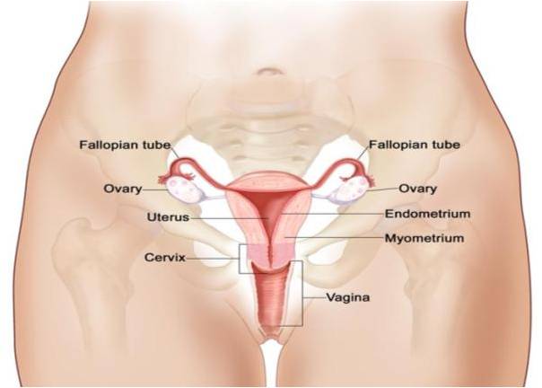
Fig. 3 (Copyright © 2011 National Cancer Institute); Location of the ovaries within the true pelvis
Functions of ovaries
The ovaries produce the eggs/ova by a process called oogenesis, which are release during ovulation. They also produce the female sex hormone called the estrogen and progesterone. The estrogen causes the thickening of the uterus and vagina during the early phase of menstruation. It also play role in the development of the female sex characters, such as breast and pubic hair.
While the progesterone is very essential for the successful implantation of the fertilize eggs and for the maintenance of the pregnancy.
**************************************************************************************************************
(Copyright © 2011 by U. Bala)
(Copyright © 2011 by U. Bala)
