Diagnosis for Ovarian cancer
Ovarian cancer is difficult to diagnose because symptoms are easily confused with those of other diseases and with the fact that, there are no perfect screening tools to identify the disease at the early stage. This cause a delay in the diagnosis of the disease, and in any case, some tests that examine the ovaries, pelvic area, blood and the ovary tissues are used to find and diagnose ovarian cancer.
Pelvic examination
This is a clinical procedure that examines the female reproductive organs, which include the vagina, cervix, uterus, fallopian tube, ovaries and the rectum for any mass. It involved inserting one or two lubricated, gloved fingers of one hand into the vagina and the other hand is place over the abdomen to feel the size and shape and position of the uterus and ovaries (fig. 7)
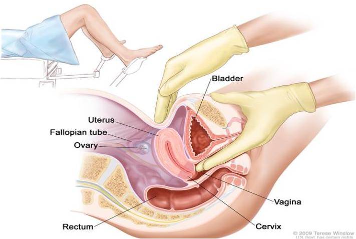
Fig. 7 (Copyright © 2011 National cancer Institute); Pelvic examination to detect the presence of mass within the pelvic area
Ultrasound Examination
This procedure requires the use of sound waves on the internal tissues or organs and produce echoes, which in turn form images (sonogram) of the body tissues. These images can be look at for any abnormal mass in the area. There are two forms of ultrasound for ovarian cancer diagnosis,
Tran-abdominal ultrasound; This is when the ultrasound transducer is passed over the surface of the abdomen. The transducer is connected to the computer and all images produced can be seen on the computer screen. Changes in the ovary that are suggestive to the tumour such as enlargement can be detected.
Tran-vaginal ultrasound; In this technique, the ultrasound probe is inserted into vagina and is gently moved to show different organs within the pelvic area internally. Also images of all structures will be display on the screen. This technique allows for better visualizations of the changes in the ovary, but is not good in distinguishing between the benign and malignant masses (fig. 8)
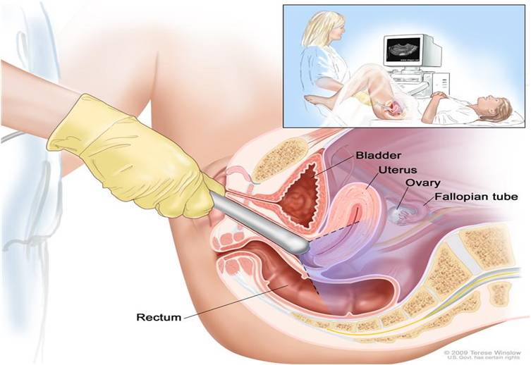
Fig. 8 (Copyright © 2011 National cancer Institute); Trans-vaginal ultrasound
CA-125 assay
The CA-125 is a serum tumour maker which is released into the blood stream. An elevated level of CA-125 is considered as a sign of ovarian cancer. Studies show that, about 80 % of the women with advance ovarian cancer have elevated CA-125 level. But other conditions such as endometriosis, pelvic inflammatory diseases, benign cyst, fibroids, and other malignancies are associated with an elevated level of CA-125. Hence, an elevated level of CA-125 alone is neither sufficiently sensitive nor sufficiently specific to be diagnosis.
Computed Tomography Scan (CT Scan)
This is an imaging technique that produces a series of detailed images of areas of body, usually taken at different angles. It involved injecting of a dye (contrast medium) into a vein to assist in clear visualization and images of the tissue or organ. The images are produced by a computer connected to an x-ray machine (fig. 9)
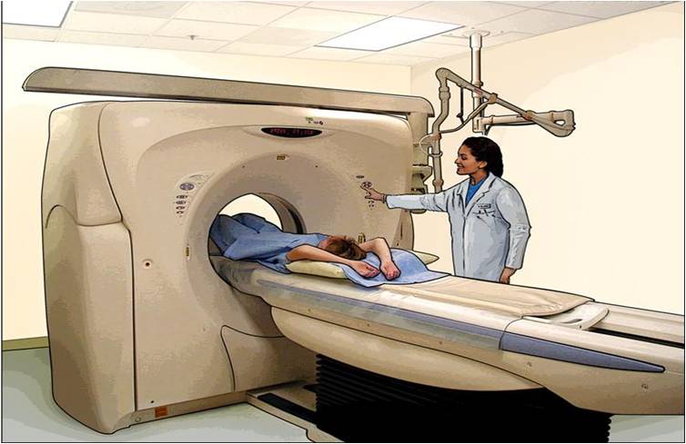
Fig. 9 (Copyright © 2011 National cancer Institute); Computed Tomography scan used in diagnosing cancer
Biopsy
This is the removal of cells or tissues for microscopic viewing (examination) to check for any signs of cancer. In this case, a procedure called laparaotomy (a surgical incision made in the wall of the abdomen) is applied to remove the tissue. During this procedure, cyst or other suspicious area must be removed and biopsied. If the lesion is cancerous, then, a process called surgical staging is done to find out how far the cancer has spread.
This is the removal of cells or tissues for microscopic viewing (examination) to check for any signs of cancer. In this case, a procedure called laparaotomy (a surgical incision made in the wall of the abdomen) is applied to remove the tissue. During this procedure, cyst or other suspicious area must be removed and biopsied. If the lesion is cancerous, then, a process called surgical staging is done to find out how far the cancer has spread.
*************************************************************************************************************
(Copyright © 2011 by U. Bala)
(Copyright © 2011 by U. Bala)
